← Back to Class 9 Science Notes
This chapter helps students understand cell structure, types of cells (prokaryotic and eukaryotic), and important cell organelles like the nucleus, mitochondria, and chloroplast. It also explains how cells perform essential life processes such as growth, respiration, and reproduction, making it a fundamental topic in Class 9 Science.

Cell The Building Block of Life Class 9 Notes
Scientists believe that life may have originated in a hot water environment rather than in deep oceans. One example is the hot springs of Puga Valley, where the conditions are similar to early earth about 3.5 billion years ago.
These hot springs have tiny living organisms called thermophiles, which can survive in very high temperatures. In this spring the scientists have found calcium carbonate deposits. This deposit may have helped in two ways:
- They protect early molecules.
- They helped from the first simple cell membranes.
What is a Cell?
All living organisms are made up of cells. A cell is the smallest unit of a living thing that can do all the life activities. The cells are called the building blocks of life.
Type of organisms based on number of cells:
| Type | Meaning | Examples |
|---|---|---|
| Unicellular | Made of one cell | Amoeba, Bacteria, Yeast |
| Multicellular | Made of many cells | Plants, Fish, Humans |
Levels of Organization in Living Organisms
Living organisms are made in a step-by-step methos like Cells → Tissues → Organs → Organ Systems
Explanation
- Cells → Smallest units of life
- Tissues → Group of similar cells doing the same work
- Organs → Group of tissues working together
- Organ Systems → Group of organs working together
Example: Respiratory System
Nose + Trachea + Lungs → Respiratory System
- Nose helps in taking in air
- Trachea carries air
- Lungs help in breathing (gas exchange)
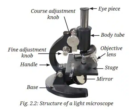
How Do We Study Cells?
Cells are very small. So we are not able to see with naked eyes. The human eye can see the object when it is bigger than 0.1 mm. Most of the cells are smaller than this, so we require a microscope to see them. In school labs we use light microscopes to magnify the objects.
Discovery of cells
In 1665, Robert Hooke was the first person to see cells using his own microscope. He saw tiny box-like structures, and he named them “cells”.
1. How to estimate the cell size
The formula used for finding the estimated cell size is: Cell Size = Field of View / Number of Cells. For example, if the field of view is 5000 µm and the number of cells is 25, then cell size – 5000/25 means the cell size will be 200 µm.
2. Magnification in a Microscope
The formula for finding magnification in a microscope is Total Magnification = Eyepiece x Objective. If the eyepiece = 10x and the objective = 10x, then the total magnification will be 10×10 = 100 µm.
Important Microscope Term
- Resolution: The resolution is used to check how clear and sharp the image looks; better resolution means better visibility.
- Contrast: It helps to make the difference between light and dark parts of the image.
- Magnification: It helps to make the object big under a microscope. It helps to make small things appear larger.
Structure of Cell
All the cells are surrounded by a thin outer boundary called the cell membrane. This membrane helps the cell to interact with its surroundings and stay alive.
Cell Membrane
The cell membrane is also called a plasma membrane. The cell membrane is a thin, flexible layer that surrounds the cell and controls the movement of substances in and out of the cell.
Functions of Cell Membrane
- The cell membrane protects the cell from damage.
- The cell membrane gives shape to the cell.
- The cell membrane controls entry and exit of substances.
- It helps the cell communicate with its environment.
Example of Cell Membrane in Lungs (Alveoli)
In the lungs, alveoli are tiny air sacs in the lungs where breathing happens. When you breathe in, the air enters your lungs; this air has oxygen. Inside the alveoli, oxygen wants to go into the blood through the cell membrane. When you breathe out, the blood has carbon dioxide. It moves from the blood into the alveoli, and then it goes out of the body.
Diffusion and Osmosis
What is diffusion?
In diffusion the particles move from high concentration to low concentration. Example,
- When you spray perfume in one corner.
- After some time the smell spreads everywhere.
Note: The particles of the perfume move from high concentration (near the spray) to low concentration (the rest of the room).
What is Osmosis?
In osmosis the water moves through a cell membrane from high to low concentrations. For example, water entered the plant from roots. The soil has more water, and root cells have less water. The water moves from soil to root through a membrane.
Difference Between Diffusion and Osmosis
| Feature | Diffusion | Osmosis |
|---|---|---|
| Meaning | Movement of particles | Movement of water |
| Membrane | Not needed | Needed |
| Example | Perfume spreading | Water entering roots |
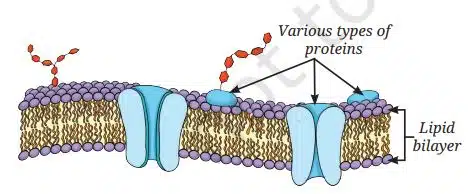
Fluid Mosaic Model of Cell Membrane
In the fluid mosaic model, the cell membrane is made of a double layer of lipids (meaning fats) with proteins scattered in it, and both lipids and proteins can move freely, which makes the membrane flexible. For example, the cell membrane is like oil with floating pieces. Where the lipids are like an oil layer and proteins are like small floating objects (doors). These proteins move and allow substances to enter and leave the cell.
Why do some cells need a cell wall?
Cells of plants, fungi and bacteria have an extra layer outside the cell membrane called the cell wall. This cell wall gives extra support and protection to the cell.
- Gives shape and strength: It helps the cell to keep its shape proper. Without the cell wall, the cells would be soft and could collapse easily.
- Protect the cell: The cell wall protects the cell from physical damage like pressure or injury and protects from substances.
- Prevents bursting: The cell takes water from the root. When too much water enters, the cell can swell. With the cell wall, it might burst.
- Support the whole organism: In plants the cell walls give strength to stems and leaves. That’s why the plants can stand straight without bones.
- Helps in communication and transport: The cell walls have tiny openings that allow movement of materials between cells, and cells can communicate between neighbouring cells.
The Cell Interior—A Coordinated Working System
The interior of the cell is a well-organized and coordinated system. It consists of three main parts.
- Plasma Membrane: It is the outer covering of the cell and is selectively permeable and controls the movement of substances in and out of the cell.
- Cytoplasm: It is a jelly-like substance inside the cell where all organelles are present and most chemical reactions occur.
- Nucleus: It is the control center of the cell. It contains genetic material (DNA) and controls all cell activities.
The cytoplasm contains organelles like mitochondria (energy production), ribosomes (protein synthesis), endoplasmic reticulum (transport), Golgi apparatus (packaging), lysosomes (waste removal), and vacuoles (storage).
All these parts work together in a coordinated way, helping the cell to function properly.
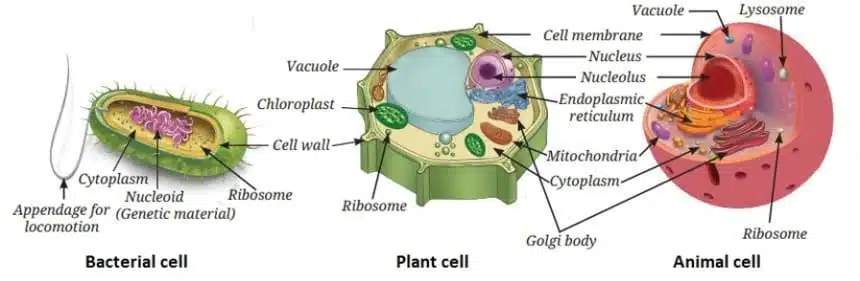
Prokaryotic vs. Eukaryotic Cells
The cells are of two main types, prokaryotic and eukaryotic.
Prokaryotic cells are simple and primitive cells. They do not have a well-defined nucleus and lack membrane-bound organelles. Their genetic material is present directly in the cytoplasm. Most activities occur in the cytoplasm. Example: Bacteria
Eukaryotic cells are complex cells. They have a well-defined nucleus and membrane-bound organelles like mitochondria, Golgi apparatus, etc. Examples: plant cells and animal cells
| Feature | Prokaryotic Cells | Eukaryotic Cells |
|---|---|---|
| Nucleus | Not well-defined | well-defined |
| Organelles | Absent | Present |
| Structure | Simple | Complex |
| Examples | Bacteria | Plants, Animals |
Why do eukaryotic cells need these organelles?
Eukaryotic cells need organelles because they perform different life processes efficiently at the same time. Each organelle has a special function, such as:
- Building new materials
- Producing energy
- Removing waste
All organelles work together like a team, making the cell function properly. Therefore, a cell is called a “tiny living factory” where every part does specific work.
Nucleus – House of coded instructions
The nucleus is the control center of the cell. It stores genetic instructions (DNA) that tell the cell what to do.
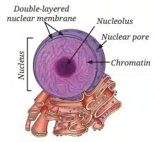
Structure of Nucleus
- Nuclear Membrane: The nuclear membrane has a double layer covering. It has pores (holes); these pores allow the materials to move in and out.
- Nucleolus: The nucleolus is a small round body inside the nucleus; it helps to make parts of ribosomes (protein makers).
- Chromatin: When the cell is not dividing, DNA exists as chromatin. It has a thread-like structure that contains DNA.
DNA, Genes, Chromosomes
- DNA: DNA contains the genetic information, or you could say DNA carries all the information of the cell.
- Genes: Genes are small parts of DNA that control traits like height, color, etc.
- Chromosomes: Chromosomes are made of DNA and proteins. It is tightly packed DNA and visible during cell division.
What Happens During Cell Division?
Cell division is the process in which one cell divides to form new cells. The steps are:
- Step 1: Chromatin become Chromosomes: DNA (loose threads) become thick and visible chromosomes.
- Step 2: Nucleus divides: The nucleus splits into two nuclei.
- Step 3: Cytoplasm divides: The rest of the cell splits.
- Step 4: Two new cells are formed: each new cell will get some genetic material (DNA).
Ribosomes — The Protein Factories
The ribosomes are very small structures in the cell that make proteins. These ribosomes are found freely in the cytoplasm and attached to the endoplasmic reticulum (RER). The functions of the ribosomes are
- Ribosomes are the sites of protein synthesis.
- They help in making proteins for growth, repair, and body function.
Endoplasmic Reticulum (ER)—Manufacturing factory
The endoplasmic reticulum (ER) is a network of membranes inside the cell that works like a manufacturing and transport system. The endoplasmic reticulum (ER) looks like a network of tubes and sacs, present in the cytoplasm and connected to the nuclear membrane.
What does ER do?
The ER helps to transport materials inside the cell like proteins, fats (lipids), and hormones (in some cells).
Types of ER
There are two types of ER.
- Rough Endoplasmic Reticulum (RER): It looks rough under an electron microscope because it has ribosomes attached to its surface and is mainly involved in protein synthesis and protein secretion (for example, in gland cells, such as pancreatic cells).
- Smooth Endoplasmic Reticulum (SER): It does not have ribosomes on its surface and therefore looks smooth. It is involved in the synthesis and storage of fats and hormones.
Golgi apparatus—The packaging and shipping centers
The Golgi apparatus is the packing and sending center of the cell. It is made of stacked, flattened sacs present in the cytoplasm and connected with the endoplasmic reticulum (ER).

What does the Golgi apparatus do?
The Golgi apparatus is just like a post office; it helps to modify proteins and fat, sorts them, packages them into small sacs called vesicles, and sends them to the different parts of the cell or outside.
Lysosomes—The clean-up system
Lysosomes are the cleaning units of the cell. It has a small sac-like structure, surrounded by a single membrane and filled with digestive enzymes.
What do lysosomes do?
Lysosomes break down and clean the waste materials and old or damaged organelles and clean the unwanted proteins, fats, and carbohydrates.
Mitochondria—The powerhouse of the cell
Mitochondria are called the “powerhouse of the cell” because they produce energy. The mitochondria are surrounded by two membranes. The outer membrane is smooth and porous, and the inner membrane is folded into finger-like projections called cristae, which increase the surface area for chemical reactions and facilitate energy production.
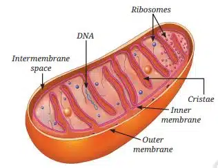
What do mitochondria do?
The mitochondria release energy by breaking down the glucose (food); this process is called cellular respiration. The energy is stored as ATP (adenosine triphosphate). ATP is the energy currency of the cell.
Plastids—centers for food synthesis in the plant cells and beyond
Plastids are special organelles found in plant cells that help in making food, storing food, and giving color to plants. Plants prepare their food by the process of photosynthesis in the presence of sunlight. A green pigment called chlorophyll, which is present in the chloroplast (a type of plastid), absorbs sunlight.
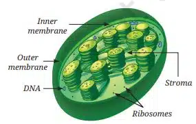
Chloroplasts are double-membrane-bound organelles, like mitochondria. Inside the chloroplast, there is a semi-fluid substance called the stroma. Within
The stroma are disc-shaped membrane structures that contain chlorophyll. Light energy is absorbed by them during photosynthesis. The sugars synthesised in this process are stored in stroma, along with the starch granules.
How do flowers, fruits, and vegetables acquire varied colors?
The different colors in plants are due to special cell organelles called “plastids.” These plastids are found in flowers, fruits, and some vegetables. This plastid contains pigments other than chlorophyll, which give color like red, orange, and yellow. These plastids are called chromoplasts.
Vacuoles—The organelles for storage and support
The vacuoles are storage sacs in the cell that store water, food (minerals and sugars), and waste. The vacuoles are surrounded by a single membrane, filled with a liquid called cell sap. In plant cells there is one large central vacuole, and in animal cells there is a small vacuole or something absent. In plants, the vacuole maintains pressure and keeps the plant firm and upright.
Why do plants wilt?
When the plant doesn’t get enough water, then the vacuole loses water and pressure, which makes the plant soft and wilted.
How Do Normal Cells Grow and Divide?
Cells help our body grow, repair, and replace old or damaged cells by dividing. The cell can grow only to a limited size. The cells divide to help heal wounds, replace dead cells, help in body growth, and form new tissue like hair or skin.
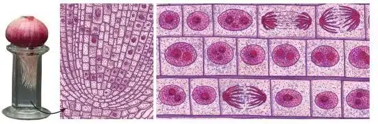
Onion Root Experiment
The root tips of the onion grow fast. Their cells are continuously dividing. Under a microscope, cells look different because they are in different stages of division.
What is Cell Division?
Cell division is the process by which new cells are formed from pre-existing cells. It allows living organisms to grow, repair damaged tissues, and reproduce. Some cells, such as skin cells, divide continuously to replace cells that are lost regularly. There are two major types of cell division — mitosis and meiosis.
Mitosis
One cell dividing into two identical cells is called mitosis. Every human begins life as a single fertilized egg. This one cell divides repeatedly to form trillions of cells in the body. Cells increase in number through mitosis, which is the most common type of cell division. Mitosis produces two genetically identical daughter cells from one parent cell. Each new cell gets the same DNA and the same number of chromosomes as the parent cell. This ensures that genetic information is largely maintained across body cells.
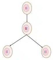
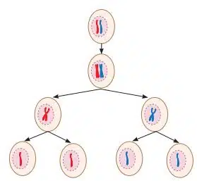
Meiosis
One cell dividing into four cells is known as meiosis. Meiosis is a type of cell division that produces gametes and occurs only in the cells of reproductive organs. Gametes produced for sexual reproduction create variations and diversity among living organisms.
Therefore, children resemble their parents but are not exactly the same. In animals, including humans, meiosis occurs only in the cells of the testes of males to produce sperm and ovaries of females to produce eggs. In plants, meiosis occurs in the anthers (male parts) to form pollen grains (that later produce sperm cells) and the ovaries (female parts) to produce egg cells. In meiosis, the parent cell divides twice, one after the other, to form four daughter cells.
Cell Theory — The Unifying Principle of Biology
Cell theory explains that all living things are made of cells and how cells are formed. According to the classical cell theory:
- All living organisms are made up of one or more cells.
- The cell is the basic unit of structure and function in living beings.
- All cells arise from pre-existing cells.
Do cells grow and reproduce forever?
No, the cells do not grow and divide forever. Every cell has a fixed life span. Cells normally grow, divide, and make a new cell. Cells do their work, and then the cells die when no longer needed. The dead cells are replaced by new cells.
Why is Control Important?
The body controls cell division carefully; if cells do not die when they should, too many cells form. If the cells die too early, the body may not function properly.
What is Contact Inhibition?
In many animal cells, they stop dividing when they touch nearby cells. This is called contact inhibition.
What happens in cancer?
The cancer cells lose control; they keep dividing continuously. This leads to the formation of tumors.
Disclaimer: The content that is present on our website is based on the NCERT Class 9 Science textbook and is provided for educational purposes only. All the content and images have been taken from Science Class 9 NCERT Textbook and CBSE Support material. Images and content shown above are the property of individual organizations and are used here for reference purposes only. To make it easy to understand, some of the content and images are generated by AI and cross-checked by the teachers.