
← Back to Class 9 Science Notes
The chapter “Tissues in Action” explains the different types of tissues found in plants and animals and their functions. Students learn how tissues help organisms carry out important activities such as support, protection, transport, and movement. This chapter also helps in understanding the structure and organization of multicellular organisms and the role of specialized tissues in maintaining life processes.
Tissues in Action Class 9 Notes
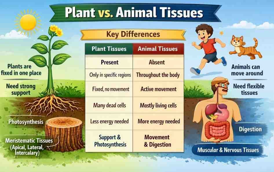
Why are Plant and Animal Tissues Different?
Plants and animals are made of cells, but their tissues are different because their lifestyles and functions are different. Plants are fixed in one place, meaning they cannot move; they need strong support to stay upright. On the other hand, animals can move from one place to another, and they need flexible tissue for movement.
Another important difference between plants and animals is their mode of nutrition. Animals have tissues that help them digest food which is obtained from different food sources, while plants have tissues that help them utilise solar energy for synthesising the food components through photosynthesis.
| Basis | Plant Tissues | Animal Tissues |
|---|---|---|
| Cell wall | Present | Absent |
| Growth | Growth occurs only in certain regions (meristematic tissues) | Growth occurs in most parts of the body |
| Movement | Plants do not move from place to place, so tissues are mostly supportive | Animals move actively, so tissues help in movement |
| Living or dead cells | Many supporting tissues are dead | Most tissues are living |
| Energy need | Less energy needed | More energy needed |
| Types of tissues | Meristematic tissues and Permanent tissues (Simple: Parenchyma, Collenchyma, Sclerenchyma; Complex: Xylem, Phloem) | Epithelial tissue, Connective tissue, Muscular tissue, and Nervous tissue |
| Function | Support, transport, storage, photosynthesis | Protection, movement, coordination, transport |
| Flexibility | Less flexible | More flexible |
Tissues for Growth in Plants
Plants grow in three different ways —
- increase in length (height of stem and depth of roots),
- increase in girth (thickness of stem), and
- regrowth after cutting the branches or grazing by animals.
This growth requires actively dividing cells that together form a tissue called meristematic tissue.
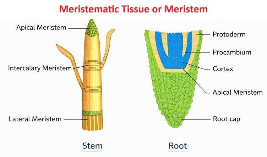
Apical meristem — How do plants grow in length?
An apical meristem is a type of meristematic tissue present at the tip of the roots and top of the shoots. It helps to increase the length (height) of the plant and is responsible for the growth of the stem upward and the growth of roots downward. This type of growth is called ‘primary growth’. Apical meristem helps the cells at the tip divide continuously. New cells are formed, while older cells enlarge and differentiate, causing the root and stem to grow in length.
Lateral meristem — How do plants grow in girth?
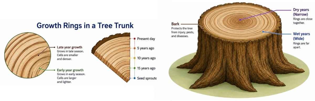
If you observe a cut tree trunk, you can see annual growth rings. These rings show the increase in girth of the plant.
This increase in girth is due to the lateral meristem, which is a type of meristematic tissue present on the sides of stems and roots. It increases the diameter of stems and roots, making the plant strong and thick. It also helps in the formation of wood (xylem) and bark (phloem).
This type of growth is called secondary growth.
Intercalary meristem — How do plants grow after being cut?
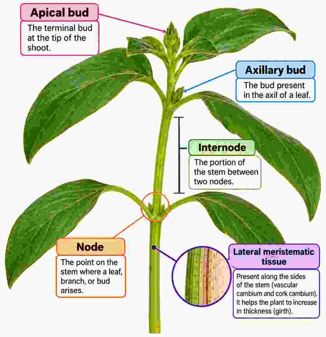
When the tip of a young stem is cut, the plant stops growing in length. However, after some time, new branches develop and the plant becomes bushy.
This happens due to the intercalary meristem, which is present at the nodes and at the base of internodes.
- A node is the point where leaves or branches arise
- An internode is the region between two nodes
The intercalary meristem helps in regrowth after cutting or damage by forming new branches and leaves, making the plant dense and bushy.
Permanent tissues
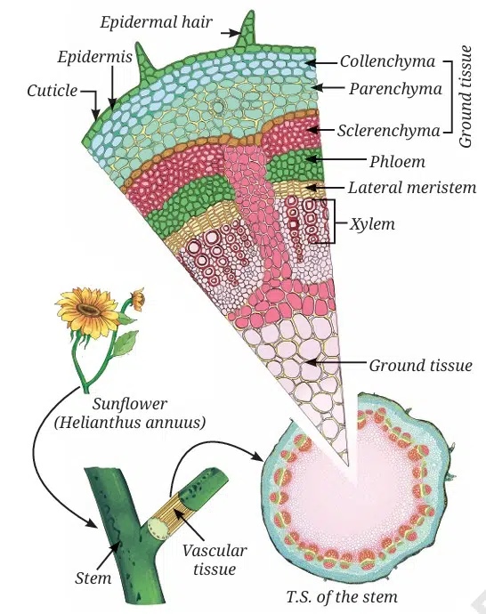
When you observe a stem, root or leaf under a microscope, you will find the cells are not all the same. They are different in shape, size and function. These different groups of specialised cells form permanent tissues. Permanent tissues can be simple (composed of only one type of cell) or complex (composed of more than one type of cell). Permanent tissues are made of cells that have lost the ability to divide and have specialised to perform specific functions.
Why are permanent tissues formed?
Permanent tissues are formed because plants need to perform many functions such as support, transport, protection, and storage.
Meristematic Tissue vs Permanent Tissue
| Feature | Meristematic Tissue | Permanent Tissue |
|---|---|---|
| Cells | Actively dividing | Do not divide |
| Shape | Small and similar | Larger and varied |
| Cell Wall | Thin | Thick or thin |
| Vacuoles | Absent or very small | Large vacuoles present |
| Intercellular Spaces | Absent | Usually present |
| Function | Helps in plant growth | Performs specific functions like storage, support, and transport |
Types of Permanent Tissues
There are two types of permanent tissues:
(i) Protective tissue — Epidermis
The epidermis is the outermost protective layer of the plant body, covering leaves, stems, and roots. It is made up of a single layer of tightly packed cells and is covered by a waxy layer called a cuticle.
Functions:
- Protects the plant from mechanical injury and harmful microorganisms
- Reduces water loss through transpiration
- In roots, epidermal cells form root hairs which increase surface area and help in the absorption of water and minerals.
(ii) Supporting tissue — Simple permanent tissues
These tissues provide strength, support, and flexibility to the plant. They are of three types:
- Parenchyma
- Collenchyma
- Sclerenchyma
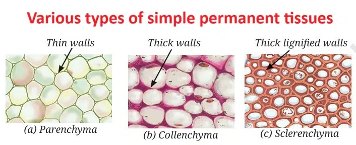
a. Parenchyma
Parenchyma consists of living cells with thin walls. These cells are loosely packed with intercellular spaces. Parenchyma mainly stores food but also performs photosynthesis in the green parts of the plants. In aquatic plants, specialised parenchyma forms air spaces, which help them float.
b. Collenchyma
Collenchyma consists of living cells with unevenly thickened cell walls at the corners due to cellulose and pectin deposition. This tissue provides support and flexibility, allowing parts of the plant like stems and tendrils to bend without breaking.
c. Sclerenchyma
Sclerenchyma cells have thick walls due to the deposition of lignin, making them hard and strong (forming the woody structure). Most of these cells are dead. This tissue is found in stems, leaf veins, and hard coverings of seeds and nuts, such as coconut husk and walnut shell.
(iii) Conducting tissues — Complex permanent tissues
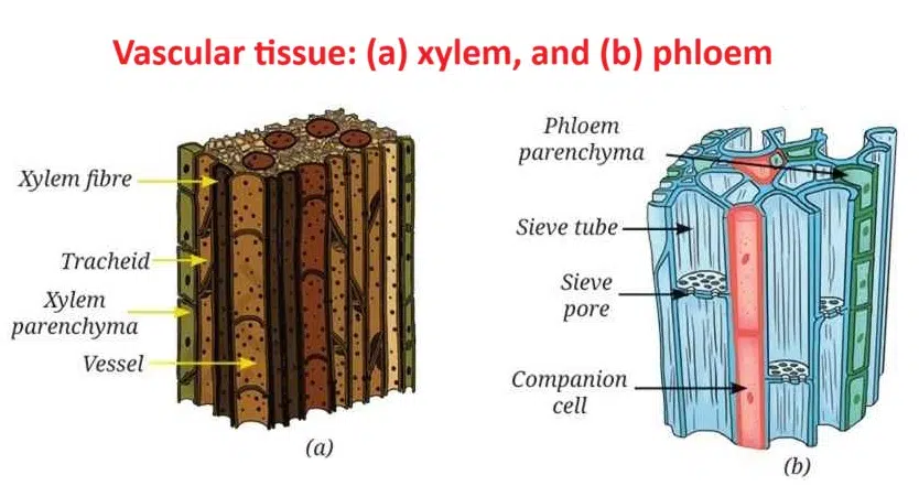
Conduction tissues in plants are specialised tissues that transport water, minerals, and food to different parts of the plant. These tissues are called complex permanent tissues because they are made up of different types of cells working together.
Types of Conducting Tissues
1. Xylem
Xylem is responsible for the transport of water and minerals from the roots to the leaves and other parts of the plant. It also provides strength to the plant.
- Tracheids and vessels are long, tubular and thick-walled.
- Most xylem cells are dead, except xylem parenchyma.
- Helps in upward movement of water
2. Phloem
Phloem transports food (sugars) prepared in the leaves to all parts of the plant.
- Mostly made up of living cells
- Sieve tubes are long tubular cells joined end-to-end with perforated walls.
- Companion cells regulate the functions of sieve tubes.
- Phloem parenchyma stores food.
Xylem vs Phloem
| Feature | Xylem | Phloem |
|---|---|---|
| Function | Transports water and minerals | Transports food |
| Direction | Mostly upward | Both upward and downward |
| Living Cells | Mostly dead | Mostly living |
| Main Components | Tracheids, vessels, fibres, parenchyma | Sieve tubes, companion cells, fibres, parenchyma |
| Provides Support | Yes | Very little |
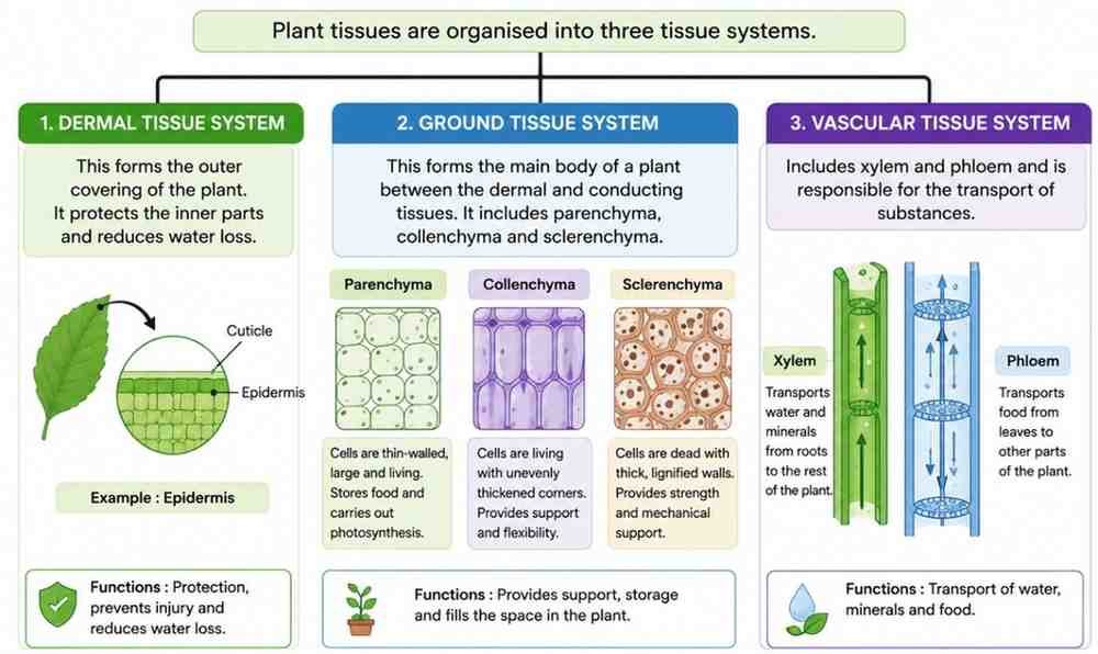
Plant tissues are organised into three tissue systems –
- Dermal tissue system: This forms the outer covering of the plant. It protects the inner parts and reduces water loss.
- Ground tissue system: This forms the main body of a plant between the dermal and conducting tissues. It includes parenchyma, collenchyma and sclerenchyma.
- Vascular tissue system: Includes xylem and phloem and is responsible for the transport of substances.
Animal Tissues
Like plants, animal cells also group together and become specialised to perform different functions. These groups of similar cells form animal tissues. Animal tissues help the body perform various activities such as movement, breathing, sensing and coordination. —
- Blink your eyes quickly.
- Clench and open your fist.
- Take a deep breath.
- Touch something warm or cold.
Epithelial tissues — Structure and functions
Epithelial tissue forms the outer covering of the body (skin) and also lines the internal organs, such as the mouth, lungs, blood vessels and intestine. It is composed of closely packed cells with very little space between them. In the types of epithelial tissues depending upon the location, they are –
- Simple squamous epithelium
- Stratified squamous epithelium
- Cuboidal epithelium
- Columnar epithelium
- Ciliated epithelium
- Glandular epithelium
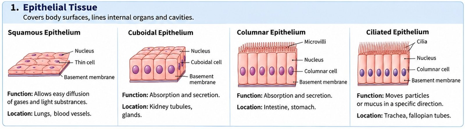
Structure of Epithelial Tissue
- Made up of closely packed cells
- Very little or no intercellular space
- Forms continuous sheets or layers
- Cells may be single-layered or multilayered depending on function.
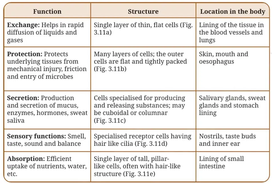
Characteristics of functionally and structurally different epithelial tissues
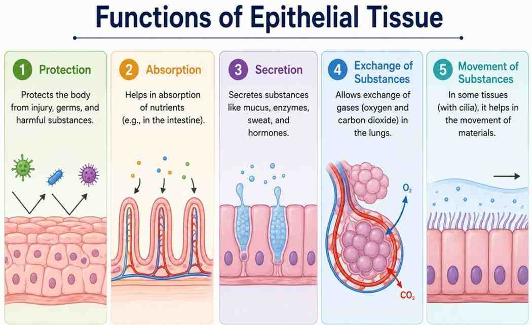
Functions of Epithelial Tissue
- Protection: Protects the body from injury, germs, and harmful substances
- Absorption: Helps in absorption of nutrients (e.g., in the intestine)
- Secretion: Secretes substances like mucus, enzymes, sweat, and hormones
- Exchange of Substances: Allows exchange of gases (oxygen and carbon dioxide) in the lungs
- Movement of Substances: In some tissues (with cilia), it helps in the movement of materials.
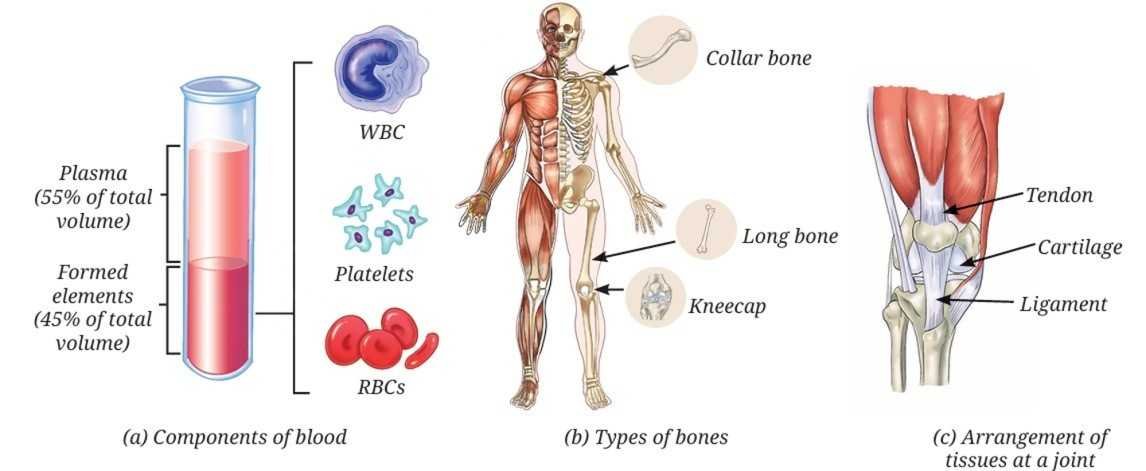
How are various parts connected in our body?
Connective tissue is a type of animal tissue that connects, supports and binds different parts of the body together. It plays an important role in maintaining the structure and coordination of the body.
Example of Connective Tissue
- Blood: It connects different parts by transporting nutrients, gases and hormones.
- Bones: The bones provide support and structure to the body.
Components of Blood and their functions
- Red Blood Cells (RBCs): RBCs contain haemoglobin; they transport oxygen to different parts of the body and give colour to the blood. The life span of RBCs is about 120 days.
- White Blood Cells (WBCs): WBCs fight infections and protect the body from diseases. Protect the body from diseases and cause inflammation at infected areas.
- Platelets: Platelets help in blood clotting and prevent excessive blood loss.
Other connective tissues are:
- Cartilage: Provides flexibility and cushions the ends of bones for shock absorption.
- Tendons: Connects muscle to bone and thus brings about movement.
- Ligaments: Connects bone to bone and provides stability, limits movement, and helps prevent dislocation.
Tendon vs Ligament
| Feature | Tendon | Ligament |
|---|---|---|
| Connects | Muscle to bone | Bone to bone |
| Flexibility | Less flexible | More flexible |
| Elasticity | Low | High |
| Function | Helps muscles move bones | Stabilizes joints |
Areolar Connective Tissue
Areolar connective tissue is a common type of loose connective tissue that binds organs, supports epithelia, and fills space between structures
Adipose Connective Tissue
Adipose connective tissue, commonly known as body fat, is a specialized loose connective tissue primarily composed of fat-storing cells called adipocytes
Can we control movement in our bodies?
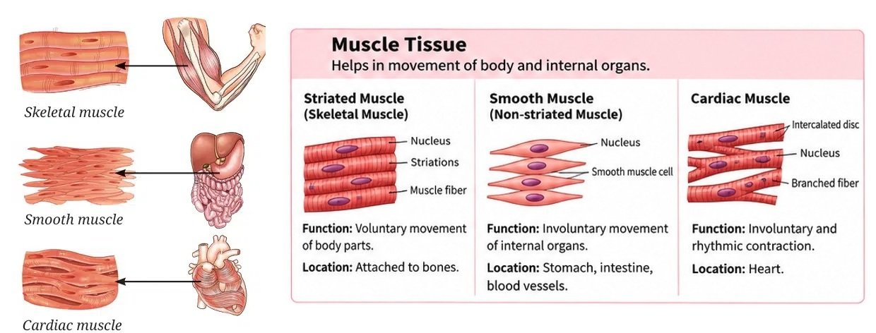
The movement in our body is controlled by muscular tissue, which works in coordination with the skeletal and nervous systems. Some movements are under our control, while others occur automatically.
Types of movements
- Voluntary Movements: These movements are under our conscious control, such as running, writing and lifting objects. The movement is controlled by skeletal muscles, attached to bones. Skeletal muscle fibres are long, cylindrical, multinucleate, and striated, helping in body movement and locomotion.
- Involuntary Movements: This movement occurs without conscious control, such as movement of food in the intestine and beating of the heart. There are two types of muscles responsible:
- Smooth Muscles: These muscles are found in the stomach, intestines and other internal organs. Their cells are spindle-shaped, have a single nucleus and lack striations. They help in slow, continuous movements like digestion.
- Cardiac Muscles: Cardiac muscles are only found in the heart. Their fibres are cylindrical and branched with a single nucleus and have faint striations. Cardiac muscles work tirelessly and rhythmically, enabling the heart to beat throughout life without fatigue.
Voluntary vs Involuntary Muscle
| Feature | Voluntary Muscle | Involuntary Muscle |
|---|---|---|
| Control | Under conscious control | Not under conscious control |
| Location | Attached to bones | Internal organs |
| Appearance | Striated | Smooth muscles are non-striated; cardiac muscles are striated. |
| Fatigue | Tires quickly | Works continuously without tiring easily |
| Example | Arm and leg muscles | Stomach and intestine muscles |
How does the body sense, communicate and respond?
Nervous tissue is responsible for control and coordination in the body. It helps the body to sense stimuli, process information, and respond quickly to changes in the environment. A neuron has three main parts: the cell body (with nucleus), dendrites (receive signals), and an axon (transmits signals to other cells).
Role of Nervous Tissue
- Controls body activities and responses
- Helps in sensing stimuli like heat, pain, and sound
- Coordinates actions such as movement and reflexes
- Works with muscles to produce responses
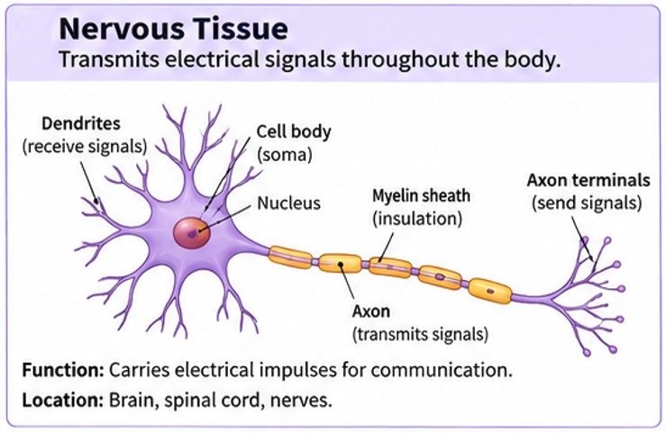
Parts of Nervous Tissue
- CELL BODY
- DENTITES
- AXON
- MYELIN SHEATH
- NODE OF RANVIER
- AXON TERMINAL END
Gap called synapse :- the specialized junction or tiny gap where a nerve cell (neuron) passes an electrical or chemical signal to another cell, such as another neuron, a muscle cell, or a gland.
The Musculoskeletal System
The musculoskeletal system is made up of bones, muscles, joints, cartilage, tendons and ligaments. This system helps us stand upright, move, maintain posture and protect delicate organs. The adult human skeleton makes up about 12–15% of body weight.
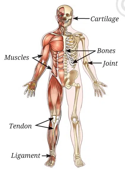
Components of the Musculoskeletal System
- Bones: Provide structure and support, and protect internal organs
- Muscles: Help in movement by contracting and relaxing.
- Joints: Connect bones and allow movement
- Cartilage: Provides flexibility and reduces friction at joints
- Tendons: Connect muscles to bones
- Ligaments: Connect bones to bones and provide stability.
Types of Joints
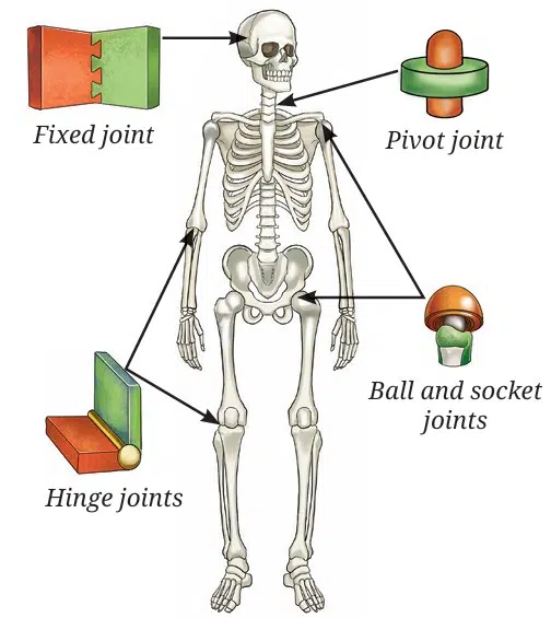
- Ball and socket joint: The shoulder joint allows free movement of the arm. This is because the rounded top of the upper arm bone fits into a shallow hollow of the shoulder bone, forming the ball and socket joint.
- Hinge joint: Unlike the shoulder, the elbow bends and straightens in one direction only like a door hinge. This type of joint is called a hinge joint. A similar hinge joint is present in the knee, where a small bone called the kneecap protects the joint.
- Pivot joint: Now, try shaking your head ‘no’. Place your hand at the back of your neck and feel the gentle movement. How does the neck move so freely? The skull is connected to the backbone through a pivot joint, which allows the head to move side to side like a doorknob turning in its socket.
- Fixed joints: The skull is a hard case of flat bones joined together to protect the brain, eyes and ears. The bones of the skull are connected by fixed joints, which means the bones of the skull cannot move. This keeps the brain safe even when the body moves.
Skeletal System
The skeletal system is a framework of bones that provides strength, support, and protection to the body. It forms the basic structure of the body and helps in movement and posture.
Main Parts of the Skeletal System
- Skull: It protects the brain and forms the structure of the face.
- Vertebral Column (Backbone/Spine): Extends from the base of the skull, Made up of small bones called vertebrae and supports the body and helps us stand upright.
The rib cage consists of 12 pairs of ribs and protects vital organs like the heart and lungs. Ribs are attached to the spine at the back and to the sternum in front, connected by flexible cartilage. This allows expansion and contraction during breathing.
Quick Revision Table
| Tissue | Living/Dead | Main Function | Example |
|---|---|---|---|
| Parenchyma | Living | Storage, Photosynthesis | Potato |
| Collenchyma | Living | Flexibility and support | Leaf stalk |
| Sclerenchyma | Dead | Mechanical strength | Coconut husk |
| Xylem | Mostly Dead | Water and mineral transport | Stem |
| Phloem | Mostly Living | Food transport | Leaf |
Exam Tips
Remember these easy tricks for exams:
- Xylem -> Water (Xylem carries water and minerals from roots to the rest of the plant.)
- Phloem -> Food (Phloem carries food prepared in the leaves to all parts of the plant.)
- Parenchyma -> Living (Thin-walled living cells and Stores food and performs photosynthesis.)
- Collenchyma -> Flexible (Gives support with flexibility and Found in young stems and leaf stalks.)
- Sclerenchyma -> Dead (Thick, lignified dead cells. and Provides hardness and mechanical strength.)
- Tendon -> Muscle to Bone
- Ligament -> Bone to Bone
- Apical -> Height (Increases the length (height) of roots and shoots.)
- Lateral -> Girth (Increases the thickness (girth) of stems and roots.)
- Intercalary -> Regrowth (Helps plants grow again after cutting or grazing.)
Disclaimer: The content that is present on our website is based on the NCERT Class 9 Science textbook and is provided for educational purposes only. All the content and images have been taken from Science Class 9 NCERT Textbook and CBSE Support material. Images and content shown above are the property of individual organizations and are used here for reference purposes only. To make it easy to understand, some of the content and images are generated by AI and cross-checked by the teachers.
i love this website this is really helpful for me for my last min studies
Nice website 👍👍
These notes are really helpful for me. I am a private tution teacher.
I am also a govt teacher, and this notes is really nice cover every topics..
Very helpful for students..
Thank you…Sir.